But: Détection et quantification de l’ADN ou de l’ARN ou des protéines cibles.
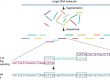
Différence avec Southern Blot Northern Blot : dépôt des échantillons directement sur une membrane de nitrocellulose, sans séparation préalable.
(+) Le dot est bien sur plus rapide est de nature a etre utiliser en routine dans des aplication medicale ou industrielle.
(-) Il donne un résultat de type présence absence. Il ne donne pas des informations sur la taille de la molécule détectée.
(-) Si deux molécules de taille différentes sont détectées, on aura un seul signal.
Principe:
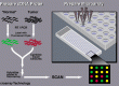
Dépôt des échantillons sur une membrane sous forme de gouttes.
Détection: on utilise deux sondes (ADN ou ARN marquées pour la détection de l’ADN ou l’ARN. Des Anticorps pour la détection des protéines.)
- sonde 1: contre un gène dont on connait le nombre de copies dans le génome, ou contre un ARN ou protéine dont la quantité de est stable durant les différentes condition testées. Cette sonde servira d’un témoin positif et de témoin de charge pour pouvoir comparer les bandes de la sonde 2.
- sonde 2 : contre un gène, un ARN ou une protéine d’intérêt.
Analyse: A l’oeil nu, ou par par le logiciel ImageJ qui permet de calculer l’intensité de chaque dot sur la membrane.

[spoiler title =”Protocole”]
Reagents
TBS:
20 mM Tris-HCl
150 mM NaCl
pH 7.5
TBS-T:
0.05% Tween20 in TBS
BSA/TBS-T:
0.1% BSA in TBS-T
Nitrocellulose membrane (BIO-RAD, Trans-Blot, etc).
Procedure
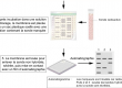
1. Have nitrocellulose membrane ready, draw grid by pencil to indicate the region you are going to blot (see below).
2. Using narrow-mouth pipette tip, spot 2 μl of samples onto the nitrocellulose membrane at the center of the grid.
Minimize the area that the solution penetrates (usually 3-4 mm diam.) by applying it slowly.
3. Let the membrane dry.
4. Block non-specific sites by soaking in 5% BSA in TBS-T (0.5-1 hr, RT). Use 10cm Petri Dish for reaction chamber.
5. Incubate with primary antibody (0.1-10 μg/ml for purified antibody, 1:1000 to 1:100000 dilution for antisera, 1:100 to
1:10000 for hybridoma supernatant) dissolved in BSA/TBS-T for 30 min at RT.
6. Wash three times with TBS-T (3 x 5 min).
7. Incubate with secondary antibody conjugated with HRP (for optimum dilution, follow the manufacturer’s
recommendation) for 30 min at RT.
8. Wash three times with TBS-T (15 min x 1, 5 min x 2), then once with TBS (5 min).
9. Incubate with ECL reagent for 1 min, cover with Saran-wrap (remove excessive solution from the surface), and expose
X-ray film in the dark room. Try several different lengths of exposure.
10. Compare the signal from your unknown sample to that of standard and estimate the concentration.
[/spoiler]